
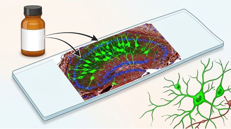
A calretinin signal can look convincing and still raise an important question: is it really calretinin?
When working with rat samples, choosing an antibody is only the starting point. The challenge is showing that the signal is specific, reproducible, and suitable for the assay. An anti-Rat Calretinin polyclonal antibody may be useful for research, but its performance should be evaluated against the needs of the experiment rather than judged solely by signal strength.
So, where should validation begin?
A structured process can reduce misleading results. Whether the goal is protein detection or tissue localization, validation helps make experimental findings easier to interpret.
Here is a practical framework for evaluating performance.
Begin With the Question, Not the Antibody
What does the experiment need to demonstrate?
That question should guide validation from the beginning. Western blotting, immunohistochemistry, immunofluorescence, and other techniques do not measure the target in exactly the same way. An antibody that performs well in one application may require different conditions in another.
Before testing, review the product information for species reactivity, applications, and sample compatibility. This helps establish a reasonable starting point instead of changing several variables at once.
Create Evidence With the Right Controls
Controls provide the context needed to judge an antibody signal. Without them, a visible result can be difficult to interpret.
A validation setup may include:
- A positive rat sample expected to contain calretinin.
- A negative sample with little or no expected target expression.
- A no-primary-antibody control to identify signal caused by secondary detection components.
- Technical or biological replicates to assess consistency.
- A second method, when practical, to provide independent evidence.
Controls should be planned before interpreting the result.
Look Beyond a Strong Signal
A bright signal is not automatically a specific signal.
For western blotting, examine whether the major band appears near the expected molecular weight and whether additional bands are present. They should prompt investigation of concentration, sample preparation, blocking, washing, and detection conditions.
For tissue or cell-based assays, examine the location and pattern of staining. Does it match the expected biology of the sample? Is the background low enough to distinguish the target from surrounding material?
These questions can be more informative than simply asking whether staining is visible.
Optimize One Variable at a Time
What should you change when the first result is weak or noisy? Avoid changing everything simultaneously, because it becomes difficult to identify which adjustment affected the outcome.
Start with the recommended antibody dilution and test a small range around it when appropriate. Then consider variables such as incubation time, blocking conditions, sample preparation, and washing.
When evaluating an anti-Rat Calretinin Polyclonal Antibody, the goal is usually a balance between adequate target detection and acceptable background. Increasing the antibody concentration may strengthen a weak signal, but it can also increase nonspecific staining. Optimization helps identify a working condition.
Compare Results Across Samples
Validation becomes more meaningful when results are compared rather than viewed in isolation.
Test samples with different expected levels of calretinin. A useful pattern should generally correspond with the biological expectations for those samples.
For example, if a positive control gives a clear signal while a negative control remains low, that provides stronger evidence than a positive result alone. Repeating the experiment can also show whether the pattern remains consistent.
Use Independent Evidence When Possible
Can one assay provide enough evidence? Sometimes, but independent confirmation can make the conclusion stronger.
An antibody used for Western blotting might be assessed alongside an imaging-based method, provided the experimental design supports that comparison. Different techniques have different sources of error, so agreement between methods can help distinguish genuine target detection from an assay-specific artifact.
Not every project needs multiple techniques.
Know What Can Affect the Result
Sample quality, fixation, antigen accessibility, protein extraction, reagent handling, and detection systems can all influence the final result.
Unexpected findings should not immediately be attributed to the antibody. Review the complete workflow and controls first. A technical issue elsewhere may produce a pattern that resembles poor specificity.
Keeping detailed records also helps. Note the antibody lot, dilution, incubation conditions, sample preparation, and detection settings so conditions can be reproduced.
Decide When the Evidence Is Sufficient
When is validation complete? There is no universal number of tests that applies to every antibody or application. Instead, consider whether the available evidence answers the important questions.
A practical validation record should establish that:
- The expected positive sample produces a suitable signal.
- Negative and reagent controls show acceptable background.
- Results can be reproduced under the selected conditions.
- The observed pattern is consistent with expected calretinin biology.
- Any limitations or unexpected findings have been documented.
Turn Validation Into a Repeatable Workflow
For researchers using an anti-Rat Calretinin Polyclonal Antibody, successful validation is ultimately about evidence. Start with the experimental question, use controls that challenge the result, optimize conditions carefully, and confirm findings when appropriate. This approach can help produce cleaner data and give researchers greater confidence when interpreting calretinin detection in rat samples.